


Congenital glaucoma
However, of all children who have lost their sight completely, one in ten did so specifically as a result of congenital glaucoma.
Types of congenital glaucoma
Two types of congenital glaucoma are distinguished:
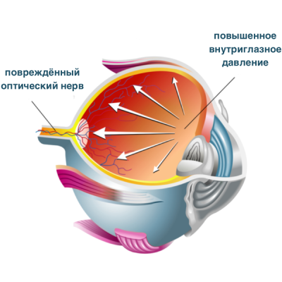
These factors cause abnormal formation of the anterior chamber angle, excessive production of aqueous humour with impaired drainage, leading to elevated intraocular pressure. In the period from birth to one year of age, the tissues of the eyeball may actively stretch under the influence of rising intraocular pressure, resulting in enlargement of the eyeball itself, and stretching and enlargement of the corneal diameter.
Congenital glaucoma is also classified as follows:
By intraocular pressure (IOP):
- Normal — no corneal oedema; Maklakov pressure up to 23 mmHg
- Moderately elevated — Maklakov pressure 23–30 mmHg; no corneal oedema or mild intermittent oedema
- High — Maklakov pressure 31–40 mmHg; persistent corneal oedema, band-shaped and cloud-like corneal opacities
- Benign — clinical manifestations develop slowly, most commonly between 1 and 2 years of age; minimal enlargement of the eye (intermediate form between infantile and juvenile congenital glaucoma)
By clinical course:
- Malignant — the final stages are already present at birth, or buphthalmos progresses rapidly in the first 1–2 months of life
- Typical — the condition develops prominently from the age of 3–4 months, with photophobia, blepharospasm, and lacrimation, indicating a fairly advanced process
- Benign — clinical manifestations develop slowly, most often between 1 and 2 years of age; minimal enlargement of the eye
- Abortive — intraocular pressure normalises spontaneously and progression ceases
By disease dynamics:
- Stable (cessation of pathological enlargement of the eye and deterioration of vision) — this occurs only following effective surgery
- Progressive (ongoing pathological enlargement of the eye and deterioration of vision) — invariably observed prior to surgery or post-operatively when no positive effect has been achieved (intraocular pressure remains elevated). Post-operative progression of glaucoma provides grounds for urgently considering indications for further surgical interventions
Diagnosis of congenital glaucoma
Timely and thorough ocular examination is essential for the treatment of congenital glaucoma.
At Crystal Vision, for children under 5–6 years of age with suspected congenital glaucoma, such examination is performed only under general anaesthesia, as any movement by the young patient may yield insufficiently accurate diagnostic results.
Diagnostics includes:
- Measurement of intraocular pressure
- Examination of the cornea and anterior chamber angle
- Measurement of the axial length of the eyeball
- Fundus examination and assessment of the optic disc
In children from the age of 3, we perform optical coherence tomography to clarify the degree of optic nerve damage and monitor changes over time.
Situations frequently arise in which a child is incorrectly diagnosed with congenital glaucoma on the basis of only 1–2 signs. In our clinical practice, through dynamic monitoring of children, we have on multiple occasions revised a diagnosis of congenital glaucoma to the correct diagnosis after conducting the full range of diagnostic procedures. It is important to understand that an incorrect diagnosis leads to the need for surgery which, in the absence of genuine indications, may cause irreversible damage to the optic nerve.
Causes of congenital glaucoma
The condition is based on impaired outflow of intraocular fluid, leading to elevated intraocular pressure. The causes include various pathological conditions in the mother, particularly during the first months of pregnancy (infections, toxic exposure, etc.).
Symptoms of congenital glaucoma
We would like to draw parents' attention to the fact that in approximately 55% of children, the first signs of glaucoma appear at an early age (from the first days of life to 5–6 years).
If you notice corneal clouding in your child's eyes, or if at birth the eyes appear large and "expressive," these may be the first indicators of congenital glaucoma. In 75% of cases it develops in both eyes.
The main symptoms of congenital glaucoma also include lacrimation, narrowing of the palpebral fissure, and photophobia.
Congenital glaucoma frequently coexists with concurrent defects in other systems and organs (microcephaly, cardiac defects, deafness, phakomatoses, etc.) as well as within the eye itself (microcornea, aniridia, cataract, etc.).
It is important for parents to know that congenital and childhood glaucoma in the majority of children does not cause complaints.
At advanced stages of glaucoma, stretching and thinning of the conjunctiva and other coats of the eyeball are observed. Complicated cataract frequently develops. In the initial stage of congenital glaucoma, the fundus is normal.
The most serious complication of congenital glaucoma is blindness, resulting from impaired blood supply to the optic disc.
Treatment of congenital glaucoma
Congenital glaucoma most commonly requires surgical intervention. At Crystal Vision, following thorough diagnostics, the surgical method is selected individually, taking into account all of the child's characteristics — including the stage of the condition and the degree of involvement of ocular structures.
Surgical treatment may include:
- Drainage of the anterior chamber angle. This is a unique technique which in most cases achieves stabilisation of the glaucomatous process and normalisation of intraocular pressure. Together with our American colleagues, we believe this intervention allows the cause of glaucoma in children to be fully eliminated, often in a single surgical procedure.
- Trabeculotomy. Used when corneal clouding is pronounced — the trabeculum (a section of the anterior chamber angle) is incised through a scleral incision to improve aqueous humour outflow.
- Trabeculectomy. Part of the trabeculum is removed through a scleral incision, creating a drainage pathway for fluid to flow beneath the conjunctiva, thereby reducing intraocular pressure.
- Goniotomy. Performed at early stages of glaucoma. A section of the anterior chamber angle is incised through the cornea; the effectiveness of this procedure in such cases is 85%.
- Combined approach (trabeculotomy + trabeculectomy)
All surgical procedures in children require general anaesthesia!
Prognosis for glaucoma treatment in children
The prognosis for congenital glaucoma depends on the age at which treatment begins and on concurrent ophthalmological disorders. According to our statistics, the most favourable prognosis is when treatment is initiated between 2 and 12 months of age — at which point the highly qualified ophthalmologists and ophthalmic surgeons at Crystal Vision achieve success in 90% of cases!