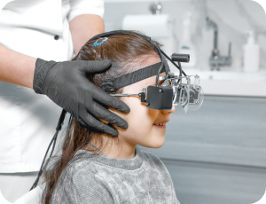
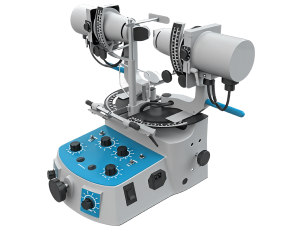
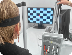
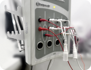
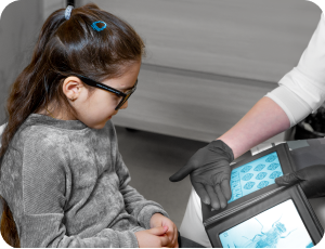
We have developed our own high diagnostics standards that allow us to:

Due to the advanced diagnostic standards developed and applied by us, the possibility of making an incorrect diagnosis is eliminated.

We guarantee that your child will be assigned the examinations that are necessary for a precise advanced diagnosis.

Equipment for diagnostics of eye diseases

Methods of treatment and diagnosis of vision

EEG

EEG
Electroencephalography (EEG) is recommended in the case of myopia, strabismus, nystagmus, amblyopia, astigmatism, and hyperopia to clarify the neurological state and revise the treatment regimen. Read more

PediaVision: remote autorefractometer

PediaVision: remote autorefractometer
At our clinic, we use the unique PediaVision Care paediatric autorefractometer — a device designed for non-contact vision diagnostics in our youngest patients. Read more

Autorefractometry

Autorefractometry
Modern ophthalmology is unthinkable without high-precision equipment. One such device is the autorefractometer — a computerised instrument that helps the doctor determine the objective parameters of your vision in just a few minutes. This procedure has become the gold standard for primary diagnostics. Read more

Accommodometry

Accommodometry
We frequently discuss the importance of accommodation in treating children with myopia, since maintaining a high level of accommodative reserve is essential to preventing progression. Read more

Gazelab video-oculograph

Gazelab video-oculograph
Examination using the Gazelab device — developed by Professor Igor Aznauryan together with the Crystal Vision team of specialists — allows precise determination of the angle of strabismus under natural conditions, in all directions of gaze (upward, downward, into the distance, etc.). This is essential for selecting the most effective treatment approach, calibrating therapeutic devices, and determining the patient management strategy. Read more

Assessment of binocular functions and cyclotropia

Assessment of binocular functions and cyclotropia
The human eyes perform a wide range of movements in different planes, the primary ones being horizontal and vertical. Read more

Visual evoked potentials

Visual evoked potentials
This is an electrophysiological examination method that is indispensable for identifying meridional amblyopia and optic nerve atrophy, for evaluating treatment outcomes, and for the dynamic monitoring of the visual system in cases of strabismus, nystagmus, amblyopia, and optic nerve atrophy. Read more

EPR — electrophysiological examination of the eyes

EPR — electrophysiological examination of the eyes
EPR of the eyes comprises a comprehensive set of examinations designed to assess the functional state of the visual pathway and its "electrical activity." Read more

Depth perception assessment

Depth perception assessment
Depth perception is based on the fusion of images from each eye into a single percept, occurring in the higher visual centres of the brain. With depth perception, the observer is able to evaluate three-dimensional space on the basis of two-dimensional retinal images. Read more

Stereoscopic vision assessment

Stereoscopic vision assessment
Using pictures viewed through special glasses, these tests check whether the child has stereoscopic vision. The FLY test is designed for the youngest patients, using child-friendly imagery to facilitate engagement. The child is shown a picture of a fly through the glasses and asked to reach out and grab its wing — which, in the presence of stereoscopic vision, will appear three-dimensional, as if alive, like a 3D cartoon. Read more

Corneal topography

Corneal topography
It is used to assess corneal characteristics and refractive parameters (including evaluation of astigmatism), and provides information on the uniformity or irregularity of the corneal surface — enabling certain conditions to be excluded or diagnosed (such as corneal surface changes associated with contact lens wear, corneal dystrophies, epitheliopathies, and others). Read more

Computerised perimetry

Computerised perimetry
Computerised perimetry is performed to examine the visual field in order to detect defects within it and determine its boundaries.ld boundaries may be observed, and isolated areas of the visual field may be lost — giving rise to so-called "scotomas" in the viewed space. Read more

Goldman lens

Goldman lens
Examination with a Goldman lens is performed for detailed inspection of the retina and internal ocular structures (where no fundus changes are found, this is carried out once only). Read more

Magnetic resonance imaging

Magnetic resonance imaging
We perform magnetic resonance imaging (MRI) of the eyes and associated areas of the brain. Orbital MRI allows visualisation of the structure of the orbits and extraocular muscles, and identification of any pathological processes within the orbit. Read more

Optical coherence tomography

Optical coherence tomography
Optical coherence tomography (OCT) is a high-technology, non-invasive examination used to rule out occult organic lesions of the retina and optic nerve, including the nerve fibre layer. Read more

Electroencephalography

Electroencephalography
Electroencephalography (EEG) is recommended in cases of myopia, strabismus, nystagmus, amblyopia, astigmatism, and hyperopia, in order to clarify neurological status and refine the treatment programme. Read more
Instalment Payment Plans