Рус Eng
+7 (495) 185 01 13 Moscow +7 (4012) 615 610 Kaliningrad

Diagnostics

We have developed our own high diagnostics standards that allow us to:

  • make a precise diagnosis,
  • identify a disease or hidden complication at the earliest stage,
  • prescribe treatment correctly.

Due to the advanced diagnostic standards developed and applied by us, the possibility of making an incorrect diagnosis is eliminated.

We guarantee that your child will be assigned the examinations that are necessary for a precise advanced diagnosis.

Equipment for diagnostics of eye diseases

Autorefractometer
Autorefractometer

Pedia Vision Сare, no-touch diagnostics of vision in the youngest patients.

Accommodometry
Accommodometry

Accommodometry is the examination of the reserve of accommodation.

Diagnostics of binocular functions and of cyclotropia

Human eyes make a lot of movements in different planes, the main of which is considered to be horizontal and vertical.

Visual evoked potentials

Visual evoked potentials (VEPs) are performed to assess the functional state of the visual system and identify subtle specific abnormalities.

Depth vision examination

The depth vision is based on the process of merging images from each eye into a single one, which occurs in the higher visual centers of the brain.

Stereo vision examination

Diagnostics of the stereo vision is performed using 2 specially designed tests: FLY stereo test and TNO test.

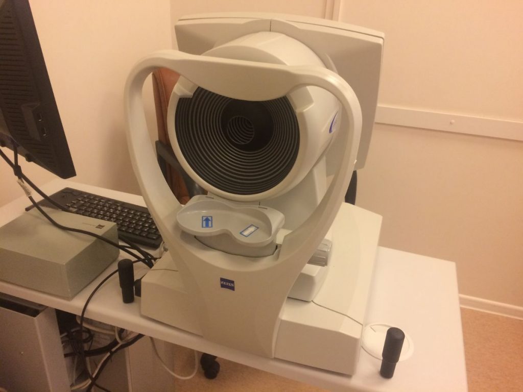
Corneal topography
Corneal topography

Corneal topography is a no-touch diagnostic procedure for assessing the condition of the cornea of the eye

Computer perimetry
Computer perimetry

Computer perimetry is used to examine the field of view in order to identify its defect and boundaries.

Goldman lens
Goldman lens

Examination using a Goldman lens is conducted for the purpose of detailed inspection of the retina and internal eye media

Magnetic resonance imaging

We perform magnetic resonance imaging (MRI) of the eyes and associated areas of the brain.

Gazelab oculograph
Gazelab oculograph

We can use this examination to determine the exact squint angle in natural conditions

Optical coherence tomography

Optical coherence tomography (OCT) is a high-tech non-invasive examination to exclude hidden organic damage to the retina and optic nerve, including the layer of nerve fibers.

Ultrasound A-scanning

Ultrasound A-scanning is performed to assess the length of the anterior-posterior segment of the eyeball, to determine its growth in dynamics, and to assess refractogenesis.

EEG
EEG

Electroencephalography (EEG) is recommended in the case of myopia, strabismus, nystagmus, amblyopia, astigmatism, and hyperopia to clarify the neurological state and revise the treatment regimen.