


Visual evoked potentials
Visual evoked potentials (VEPs) are performed to evaluate the functional state of the visual system and to detect subtle, specific abnormalities.
This is an electrophysiological examination method that is indispensable for identifying meridional amblyopia and optic nerve atrophy, for evaluating treatment outcomes, and for the dynamic monitoring of the visual system in cases of strabismus, nystagmus, amblyopia, and optic nerve atrophy.
We perform VEPs in children from 3 months of age.
In children under 2 years, VEPs are used to monitor the maturation of the visual pathway, and the examination may therefore be performed every 3 months; after the age of 2, it is carried out approximately once a year.The examination lasts between 30 minutes and 1 hour.
How to prepare your child for VEP examination:
- Wash your child's hair the evening before, without using conditioner.
- Make sure your child has had a good night's sleep.
- If your child wears glasses, be sure to bring them. If your child wears contact lenses, it is also advisable to bring them or to arrive wearing them (some examinations are best performed in contact lens correction). Do not forget to bring a lens case with solution in case the lenses need to be removed.
- If your daughter wears earrings, remove them in advance.
- If you are taking any medication regularly, take it as usual. Other medications that you take occasionally are best avoided before the examination. Inform the examining doctor of all medications you are currently taking.
- Please arrive at your appointment in good time and try to ensure your child is relaxed before the examination.
Recommendations may vary depending on the type of electrophysiological examination.
How VEP examination is performed
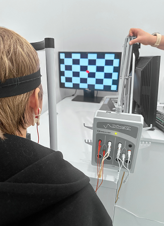
VEP is a safe and non-invasive procedure. Here is what to expect:
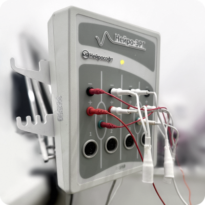
 Before the examination begins, the doctor will attach several electrodes to the earlobes and scalp. Saline solution, contact gel, or paste may be used for this purpose.
 The doctor will give instructions to you or your child about what to do during the examination. Typically, each eye is examined separately (covered using an occluder or patch).
 After the examination, the doctor will analyse the results and discuss the findings with you.
After the examination, the doctor will analyse the results and discuss the findings with you.