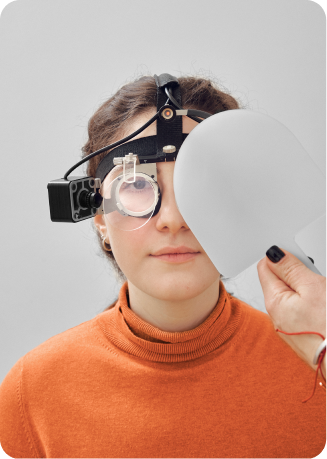


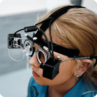
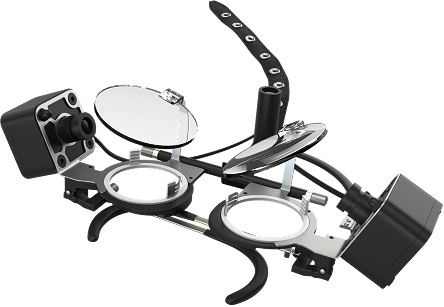
Gazelab video-oculograph
Examination using the Gazelab device — developed by Professor Igor Aznauryan together with the Crystal Vision team of specialists — allows precise determination of the angle of strabismus under natural conditions, in all directions of gaze (upward, downward, into the distance, etc.), which is essential for selecting the most effective treatment approach, calibrating therapeutic devices, and determining the patient management strategy.
In paralytic strabismus, Gazelab assesses ocular motility, which is necessary for accurate surgical planning and monitoring post-operative recovery.
This approach ensures high precision in the treatment of both strabismus and nystagmus.
A unique patented method for examining strabismus and nystagmus
- For effective treatment of strabismus
- For calibrating therapeutic devices
- For determining the management strategy for patients with strabismus
In patients with strabismus, this examination allows us to determine the precise angle of deviation under natural conditions — since what matters most is how the eyes behave in everyday life, not under provocative tests in the ophthalmologist's office.
Gazelab identifies the angle of deviation in all directions of gaze (upward, downward, into the distance, etc.).
In paralytic strabismus, the video-oculograph enables the doctor to examine ocular motility — which is critically important for accurate surgical dosing and for monitoring changes during the post-operative period.
In patients with nystagmus, nystagmography allows identification of the head position at which oscillatory movements are absent or minimal.
Measurements of nystagmus frequency and amplitude allow the doctor to monitor the progress of conservative treatment and to determine the surgical approach, where this is required.
Who can benefit
- Examination using Gazelab — an innovative method for precisely determining the angle of ocular deviation
- Magnetic resonance imaging — to clarify orbital anatomy and exclude anomalies
- Neurologist consultation — to identify possible causes of the eye condition
- Stereoscopic vision assessment and electroencephalography — for the most accurate determination of the child's neurological status
- Assessment of binocular functions — to determine whether both eyes are able to perceive a single, three-dimensional image