


Optical coherence tomography
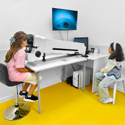
Optical coherence tomography (OCT) is a high-technology, non-invasive examination used to rule out occult organic lesions of the retina and optic nerve, including the nerve fibre layer.
Note! Pupil dilation is required for this examination and will persist for 3-4 hours.
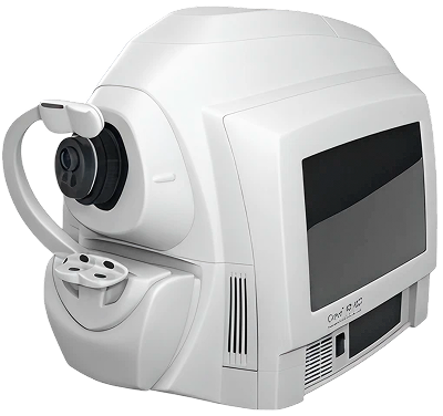
Equipment used to perform OCT
The OCT Zeiss Cirrus device is a modern optical coherence tomography (OCT) system that performs high-precision scanning of ocular structures without contact or pain. It enables detailed, layer-by-layer examination of both the posterior segment of the eye (retina and optic nerve) and the anterior segment (cornea, anterior chamber), delivering high-accuracy diagnostics at a microscopic level.
In the posterior segment of the eye, it is possible to examine the retina with assessment of its thickness and layered structure, and the macular region (the central area of vision). Additionally, the optic nerve head, the nerve fibre layer, and the ganglion cell layer are analysed — which is particularly important for the early detection of glaucoma, macular degeneration, and other retinal pathologies.
In children, using the OCT Zeiss Cirrus in the posterior segment of the eye allows the detection of congenital abnormalities of the retina and macula, as well as changes in the optic nerve, including early signs of glaucoma and atrophy. The method enables the identification of pathology at early stages and the monitoring of ocular development without invasive intervention.
In the anterior segment of the eye, it is possible to examine the cornea with measurement of its thickness and assessment of its structure, the anterior chamber (its depth and configuration), and the anterior chamber angle — which is important for the diagnosis of closed-angle glaucoma.
Related articles
One of the most common causes of reduced vision in children. In amblyopia, one eye participates very little in the process of seeing, and the brain receives visual information almost entirely from the healthy eye. It frequently develops as a complication of strabismus, hyperopia, or astigmatism.

A condition in which the fibres of the optic nerve — which transmit information from the eye to the brain — are destroyed. It manifests as reduced visual acuity and narrowing of visual fields.
Atrophy results in a portion of visual information failing to be processed by the corresponding areas of the brain, and vision deteriorates significantly.